When I reflect on the changes in medicine in the almost 50 years since I graduated from medical school, I list advanced imaging as the most significant. At the time, the only tool available to examine structures within the body nonsurgically was a plain X-ray.
On an X-ray, I could see bones and could almost always diagnose a significant bone fracture or joint disruption. In those days, when an expansive history and a detailed physical exam were not only key components of the medical curriculum but also essential skills for any physician, I could order a chest X-ray (invented 1895) on a feverish patient with a cough and see the pneumonia, which I already knew was present thanks to my stethoscope (invented 1860).
I attended medical school at the University of Virginia from 1977 to 1981, and I remember when the first head computed tomography (CT) scanner was installed. Because the state of Virginia required a certificate of need for this high-cost device, it was purchased by other means and installed in a building across the street from the hospital. It could only perform scans of the brain, and the patient had to be immobile for several minutes while the gantry rotated around their head. A room full of computers then created a very pixelated image in less than an hour. It seemed a miracle to us. In those days, we referred to the machine as an EMI scanner because EMI—the same company that produced the Beatles—was a major supplier of head CT scanners.
Before the CT scanner, the diagnosis of a brain tumor was based on a very detailed neurologic exam by a physician with vast experience in determining the probable location of the tumor by subtle abnormalities in the exam. If the odds of a tumor being the cause of the patient’s symptoms were high enough, the patient was admitted for three days and had multiple studies performed by a team of neuroradiologists.
Contrast was injected into each of the arteries serving the brain, and X-rays were performed, producing a four-vessel angiogram. After this was completed, the patient was placed in a gimbaled chair, and a dense contrast material was injected into the spinal fluid with the patient suspended upside down so that the contrast would flow into the skull and X-rays might reveal a bulge, which was unexpected in some part of the brain stem.
Finally, the patient had all their spinal fluid drained, and multiple X-rays were performed in which the surface of the brain could be seen, albeit very indistinctly. This almost unendurably painful process resulted in an image called a pneumoencephalogram. Ultimately, only opening the skull would reveal whether pathology was present and, if so, what it was.
I completed a residency in emergency medicine in 1984, when it was a brand-new specialty. There is probably no other specialty that is so focused on the art of diagnosis. Frequently, the emergency physician must evaluate a patient with abdominal pain. In the days when CT scanning was confined to the skull, a very detailed history and physical examination and a lot of experience and judgment were required to differentiate appendicitis from diverticulitis and to correctly diagnose the rapidly fatal aortic aneurysm when the majority of patients with that sort of pain were suffering from a kidney stone or something else which wasn’t immediately life-threatening.
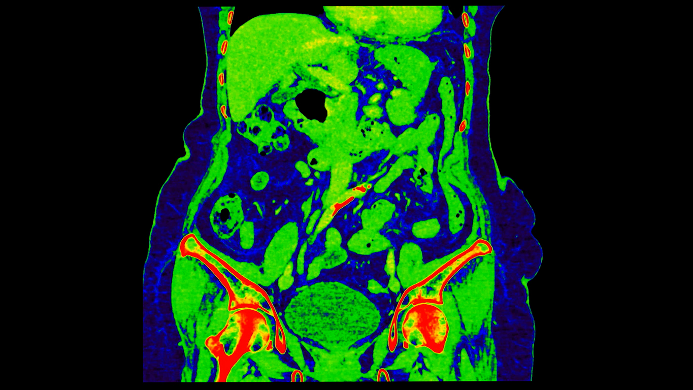
In the early 1990s, the small hospital where I worked acquired a CT scanner capable of scanning the abdomen and chest. In the Emergency Department, we seldom ordered a CT scan of the abdomen because we had developed expertise in diagnosing without relying on advanced imaging. In addition, we knew that the radiologist would complain bitterly about the “inappropriate” overuse of CT scans when they were not necessary. Interestingly, the medical necessity for a CT scan was even harder to justify after 5 PM on weekdays and weekends. However, as the scanners became faster, physicians gradually came to depend more on CT scans for patients with suspected intra-abdominal pathology.
Ultimately, CT scanning revolutionized clinical practice worldwide, and the United States adopted it with particular enthusiasm. In 1983, American providers performed an estimated 5.5 million CT procedures. By 1995, that number had surged to approximately 20 million. By 2011, CT use in the United States far outpaced that of other OECD nations, with 273.8 scans performed annually for every 1,000 people—more than twice the 16-country average of 121.7 per 1,000.
With the rapid rise in CT imaging came growing awareness of the potential for overuse and inappropriate use. This was sometimes driven by financial incentives, particularly when clinicians owned the scanner. As one physician put it to The New York Times, “If you have ownership of the machine, you’re going to want to utilize the machine.” In other cases, physicians ordered scans as part of what came to be known as defensive medicine—aggressive departures from standard practice intended to reduce legal liability.
The rapid growth in imaging not only raised costs for individual patients but also contributed to rising national health expenditures, with CT emerging as one of the fastest-growing components of Medicare spending in the early 2000s.
Perhaps most concerning was the increasing awareness of radiation-related risks, particularly for pediatric patients. As my co-authors and I noted in a 2014 paper, the use of CT scanning ultimately became a reflexive part of the workup for young people with suspected appendicitis.
That shift was especially striking given the age group most affected—10 to 19 years old—a population estimated to be ten times more sensitive to radiation than adults. We cited an Israeli study suggesting that CT use alone in pediatric patients could contribute to a 0.29% increase in future cancer mortality.
We attempted to weigh the benefits of routine scanning against its risks, noting that the risk associated with a negative appendectomy is negligible. At one institution we examined, 2,000 abdominal CT scans led to 58 appendectomies. Using BEIR V risk models, we estimated that the same 2,000 scans would result in at least one cancer death. To put that in perspective, before widespread CT use, around 23% of appendectomies turned out to be unnecessary. After the adoption of routine preoperative scanning, that figure dropped to just 1.7%. This suggests that universal scanning helped avoid 12 negative surgeries, but potentially at the cost of one radiation-induced cancer death. A trade-off of this magnitude warrants rethinking the reflexive ordering of CT in cases of suspected appendicitis. It is also a lesson that is applicable to the use of any new technology.
During the course of my career, imaging technologies revolutionized medical diagnosis and therapy, allowing clinicians to save lives that might otherwise have been lost. Yet, progress inevitably has costs and these must be acknowledged and quantified. The adoption of imaging for suspected appendicitis was associated with a growth in healthcare spending. At the clinical level, I witnessed a decline in physicians’ diagnostic abilities.
The best physicians only intervene when there is an expectation of benefit and an acceptable level of risk. That analysis requires wisdom, humility, and integrity.